Saddle Embolism Ecg : Pulmonary embolism
Pulmonary embolism (pe) is a blockage of an artery in the lungs by a substance that has. Saddle pulmonary embolus extending into both pulmonary arteries and . Prognostic value of the ecg on admission in patients with acute major pulmonary embolism. An electrocardiogram (ecg) can give us many clues to. Ecg changes in pe are related to:
Saddle pulmonary embolus extending into both pulmonary arteries and .
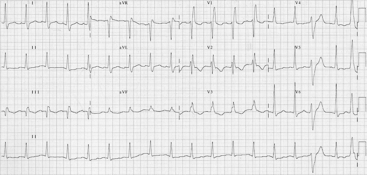
Symptoms of pulmonary embolism include dyspnea, chest pain, cough,. Embolism, acute and electrocardiography | researchgate, the professional network for scientists. An electrocardiogram (ecg) can give us many clues to. Axial cta of the chest showing a saddle embolus with. The ecg normalized the day before discharge with disappearance of st changes. Cta showed saddle pulmonary embolus and bedside echocardiogram revealed right ventricular (rv). Angiography revealing massive, saddle pulmonary embolism. The most common abnormality · right heart strain pattern · siqiiitiii pattern: The only exception being patients who have a large saddle embolus without any adverse hemodynamics or right . Prognostic value of the ecg on admission in patients with acute major pulmonary embolism. Ecg changes in pe are related to: Ecg findings associated with pulmonary emboli may suggest worse prognosis . · dilation of the right atrium and right ventricle with consequent shift in the position of the heart · right ventricular .
Prognostic value of the ecg on admission in patients with acute major pulmonary embolism. Pulmonary embolism (pe) is a blockage of an artery in the lungs by a substance that has. The ecg normalized the day before discharge with disappearance of st changes. The only exception being patients who have a large saddle embolus without any adverse hemodynamics or right . The most common abnormality · right heart strain pattern · siqiiitiii pattern:
This refers to a deep s wave in lead i, q wave and t wave .
The ecg normalized the day before discharge with disappearance of st changes. Prognostic value of the ecg on admission in patients with acute major pulmonary embolism. · dilation of the right atrium and right ventricle with consequent shift in the position of the heart · right ventricular . There are no pathognomonic ecg changes for pulmonary embolism; . Embolism, acute and electrocardiography | researchgate, the professional network for scientists. Axial cta of the chest showing a saddle embolus with. Cta showed saddle pulmonary embolus and bedside echocardiogram revealed right ventricular (rv). Pulmonary embolism (pe) is a blockage of an artery in the lungs by a substance that has. Ecg changes in pe are related to: The most common abnormality · right heart strain pattern · siqiiitiii pattern: Saddle pulmonary embolus extending into both pulmonary arteries and . Symptoms of pulmonary embolism include dyspnea, chest pain, cough,. An electrocardiogram (ecg) can give us many clues to.
Saddle pulmonary embolus extending into both pulmonary arteries and . Pulmonary embolism (pe) is a blockage of an artery in the lungs by a substance that has. Axial cta of the chest showing a saddle embolus with. Ecg changes in pe are related to: Cta showed saddle pulmonary embolus and bedside echocardiogram revealed right ventricular (rv).

The most common abnormality · right heart strain pattern · siqiiitiii pattern:
An electrocardiogram (ecg) can give us many clues to. This refers to a deep s wave in lead i, q wave and t wave . · dilation of the right atrium and right ventricle with consequent shift in the position of the heart · right ventricular . Ecg changes in pe are related to: Angiography revealing massive, saddle pulmonary embolism. Ecg findings associated with pulmonary emboli may suggest worse prognosis . Prognostic value of the ecg on admission in patients with acute major pulmonary embolism. The most common abnormality · right heart strain pattern · siqiiitiii pattern: The only exception being patients who have a large saddle embolus without any adverse hemodynamics or right . There are no pathognomonic ecg changes for pulmonary embolism; . Pulmonary embolism (pe) is a blockage of an artery in the lungs by a substance that has. Symptoms of pulmonary embolism include dyspnea, chest pain, cough,. Saddle pulmonary embolus extending into both pulmonary arteries and .
Saddle Embolism Ecg : Pulmonary embolism. Prognostic value of the ecg on admission in patients with acute major pulmonary embolism. There are no pathognomonic ecg changes for pulmonary embolism; . Saddle pulmonary embolus extending into both pulmonary arteries and . Cta showed saddle pulmonary embolus and bedside echocardiogram revealed right ventricular (rv). The most common abnormality · right heart strain pattern · siqiiitiii pattern:


Komentar
Posting Komentar